Technologies
Our dental clinic offers advanced technologies and innovative solutions to ensure precise diagnostics and individualized treatments.
From 3D scanning of the jaw (CBCT) and Digital Smile Design to modern CAD/CAM systems and high-quality prosthetic materials – every step is carefully designed to improve the experience and results of our patients.
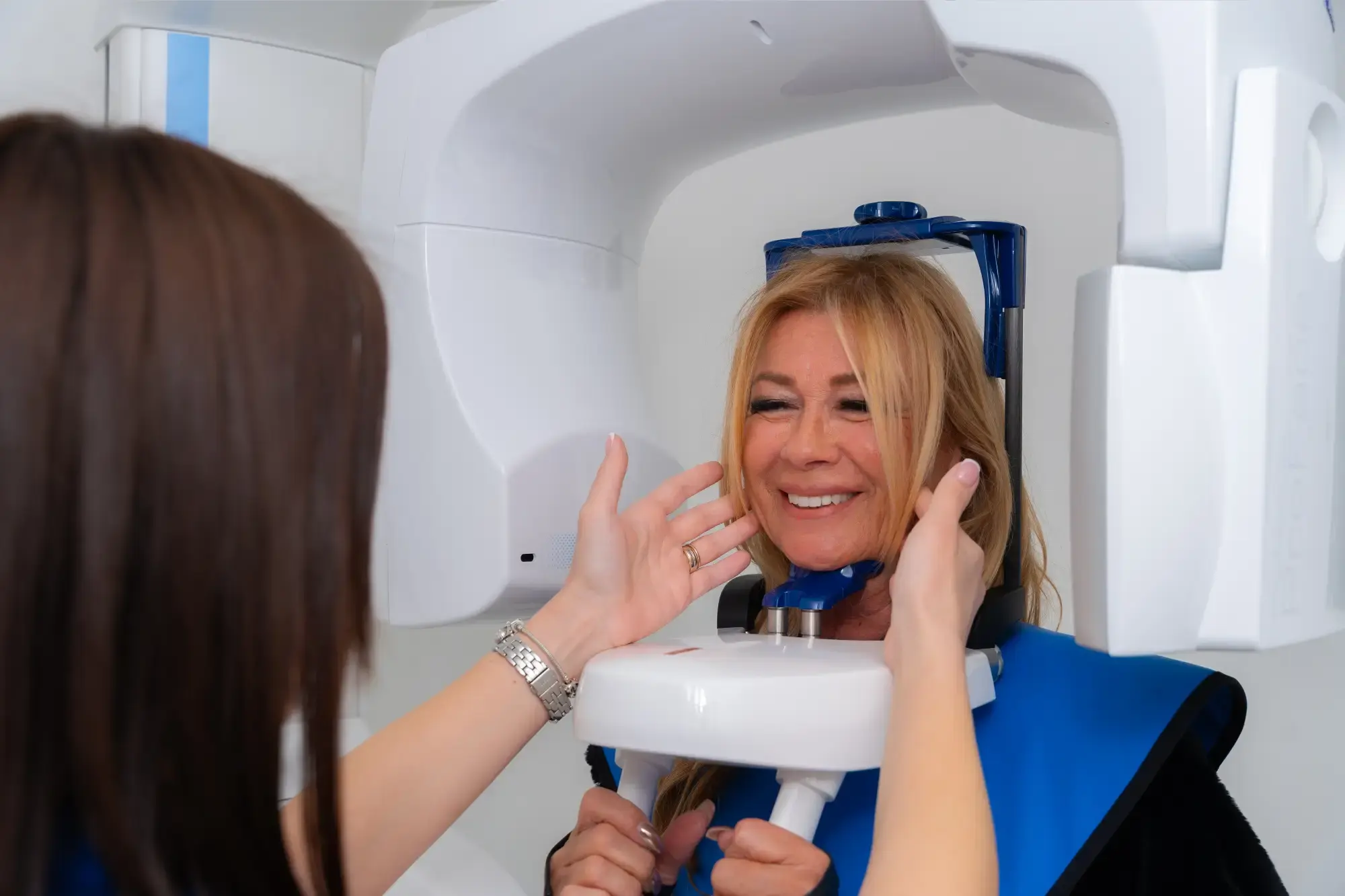
3D Jaw Imaging – 3D CBCT
What is 3D Jaw imaging?
3D jaw imaging, also known as CBCT (Cone Beam Computed Tomography), is an advanced radiological technique that provides a detailed three-dimensional visualization of the jawbones, teeth, sinuses, and surrounding anatomical structures. Unlike traditional panoramic X-rays, which offer a two-dimensional view of the jaw and teeth, CBCT scans deliver a far more accurate representation of the patient’s anatomy, enabling precise diagnosis and treatment planning.
What is 3D Jaw imaging used for?
This type of imaging is used across various dental and maxillofacial disciplines:
- Implantology – Allows for accurate planning of dental implant placement, assessment of bone thickness and density, and avoidance of nerve or sinus damage.
- Orthodontics – Assists in the analysis of tooth position, jaw structure, and identification of bite anomalies.
- Oral Surgery – Used for evaluating impacted wisdom teeth, assessing cysts, tumors, and other pathological changes.
- Endodontics (Root Canal Treatment) – Enables detailed visualization of root canal anatomy, hidden infections, and fractures.
- Periodontology – Provides insight into bone levels around teeth in advanced periodontitis cases.
- TMJ and Jaw Pain Diagnostics – Assesses the condition of the temporomandibular joint (TMJ) in patients with jaw pain, joint sounds, or dysfunction.
What are the benefits for the patient?
- Accurate diagnosis and enhanced treatment planning - The 3D view offers clinicians a detailed understanding of the condition, leading to more precise diagnoses and better-informed treatment plans with reduced risk of complications.
- Lower radiation exposure - Although it is an X-ray based technology, CBCT delivers significantly lower radiation doses compared to conventional CT, ensuring patient safety.
- Quick and painless procedure - The scan takes only 10–30 seconds, is completely painless, and requires no special preparation.
- Improved patient understanding - Patients can view their own 3D images, helping them to better comprehend their condition and the proposed treatment.
- Reduced risk during procedures - Accurate assessment of anatomical structures ensures safer execution of procedures such as implant placement, wisdom tooth extraction, or root canal treatment, minimizing complications.
2D Orthopantomographic image (OPG)
What is a 2D Orthopantomographic image?
A 2D orthopantomographic image (OPG) is a digital X-ray of the entire upper and lower jaws, including all teeth, jaw joints, and surrounding structures. This type of imaging provides a comprehensive overview of the patient's oral health and is often the first step in diagnosing dental issues.
Unlike conventional individual tooth X-rays, the OPG captures the entire oral cavity in a single image, making it extremely useful for treatment planning and assessment of the teeth and jawbone.

What is the OPG used for?
- Diagnosis of caries and tooth conditions – Helps detect cavities, old fillings, tooth fractures, and other dental issues not visible to the naked eye.
- Assessment of root and bone health – Used to identify infections, cysts, granulomas, and other pathological changes around the tooth roots and surrounding bone.
- Orthodontic treatment planning – Enables orthodontists to evaluate tooth position, root angulation, and bite irregularities before fitting braces.
- Wisdom tooth evaluation – Assists in determining the position of impacted (unerupted) wisdom teeth and their proximity to nerves and other anatomical structures.
- Planning oral surgical procedures – Essential for preparing tooth extractions, implant placements, and other surgical interventions.
- Monitoring periodontal health – Useful for analyzing bone loss and diagnosing periodontal diseases.
- Assessment of jaw joints (TMJ) – Helps in diagnosing temporomandibular joint disorders, including pain, clicking, and restricted mouth opening.
What are the benefits of OPG for the patient?
- Fast and simple diagnosis - The scan takes only a few seconds and requires no special preparation.
- Painless procedure - OPG is completely non-invasive and pain-free, with no discomfort for the patient.
- Minimal radiation exposure - Thanks to modern digital technology, patients are exposed to a very low dose of radiation, making this diagnostic method safe—even for children.
- Comprehensive view of the oral cavity - Provides the dentist with a detailed overview of overall oral health and enables early detection of potential issues.
- Accurate treatment planning - Allows the dentist to make well-informed treatment decisions, whether for fillings, prosthetics, orthodontics, or oral surgery.
Periapical dental X-ray – intraoral radiograph
What is a periapical dental X-ray?
A periapical dental X-ray (intraoral radiograph) is a diagnostic method that provides a detailed view of an individual tooth and its surrounding structures. This type of imaging is used for accurate assessment of caries, root canals, fillings, infections, and other dental problems that are not visible to the naked eye.
Unlike panoramic (OPG) or 3D CBCT imaging, a periapical X-ray focuses on a small portion of the oral cavity, offering high image resolution and allowing the dentist to make an accurate diagnosis and recommend appropriate treatment.
What is It used for?
- Detection of deep caries – Helps diagnose cavities that develop between teeth or underneath existing fillings.
- Assessment of root health – Enables the dentist to detect inflammation, granulomas, cysts, and other changes at the root apex.
- Post-endodontic treatment control – Used to verify the success of root canal therapy and ensure that no infection remains.
- Evaluation of fillings and prosthetics – Assesses the condition of fillings, crowns, and bridges, and reveals any fractures or irregularities.
- Diagnosis of periodontal disease – Allows the dentist to evaluate bone support around the tooth and assess for periodontitis.
- Pre-extraction planning – Helps evaluate root position and potential complications before tooth extraction.
- Monitoring tooth development in children – Used to assess the eruption and position of permanent teeth and detect potential orthodontic issues.
What are the benefits for the patient?
- High accuracy and detail - Provides detailedinsight into the tooth and surrounding tissues, supporting precisediagnosis.
- Minimal radiation exposure - Thanks to digital radiography, patients are exposed to a very low dose of radiation, making this method safe even for children and pregnant women (with prior consultation).
- Quick and painless imaging - The procedure takes just a few seconds, is completely painless, and requires no special preparation.
- Precise treatment planning - Enables the dentist to make informed decisions for treatment, whether it involves fillings, root canal therapy, prosthetics, or extractions.
- Reduced risk of complications - Early detection of problems allows for timely treatment and reduces the need for more complex procedures later.
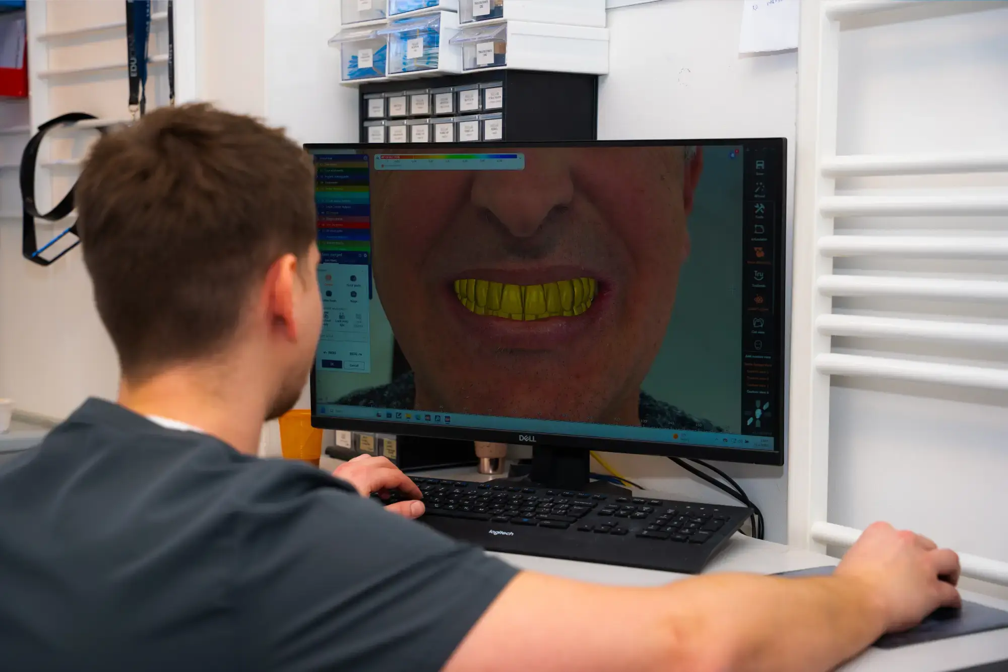
DSD – Digital smile design
What is DSD – Digital smile design?
DSD (Digital Smile Design) is an innovative dental technique that uses digital technology to precisely plan and design a smile before any treatment begins. This method allows patients to preview their future smile after aesthetic or functional procedures, giving them confidence and peace of mind throughout the transformation process.
Digital Smile Design combines high-resolution photography, video analysis, and 3D scanning to create a customized treatment plan tailored to the patient’s facial anatomy and personal preferences. The goal is a natural, harmonious, and aesthetically perfect smile that complements the patient’s unique features.
What is It used for?
- Aesthetic dentistry – Planning for teeth whitening, veneers, aesthetic fillings, and ceramic crowns.
- Smile makeover – Ideal for patients seeking a complete smile transformation.
- Orthodontic therapy – Helps visualize the final outcome before starting treatment with braces or aligners.
- Prosthetics and implantology – Enables precise planning of crowns, bridges, and implants for a natural look and proper function.
- Facial and dental structure analysis – Ensures the new smile is in harmony with the patient’s facial lines, lips, and expressions.
What are the benefits for the patient?
- Preview of the final smile - Digital simulation lets the patient see the expected outcome before starting any procedures.
- Personalized and precise treatment planning - Each smile is designed according to the facial proportions and wishes of the patient for a harmonious, natural result.
- Greater confidence and satisfaction - Patients take part in the design process and can suggest changes before treatment begins.
- Minimally invasive approach - Precise planning helps avoid unnecessary interventions and preserves natural tooth structure.
- Function meets aesthetics - Beyond aesthetics, DSD improves bite function, supporting better chewing and speech.
What does the DSD process look like?
- Photography and scanning – Digital photos and 3D scans of the oral cavity are taken.
- Smile analysis and design – Using software tools, the dentist creates a digital smile simulation.
- Patient presentation – The proposed smile is shown to the patient, who can give feedback.
- Treatment planning and production – Once approved, veneers, crowns, or other restorations are created based on the DSD plan.
- Final result – The patient receives a fully personalized smile tailored to their features and expectations.
DENTAL LABORATORY
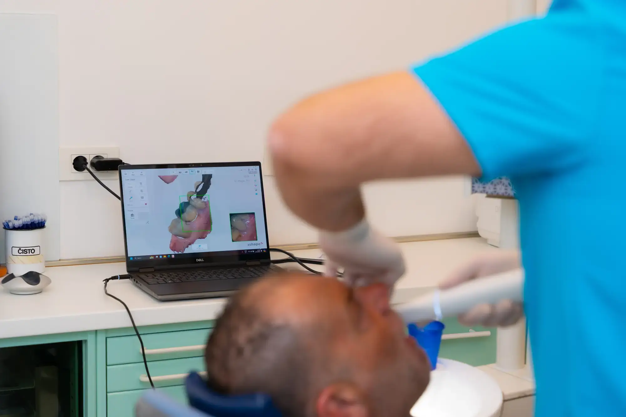
What is the Straumann digital scanner?
The Straumann digital scanner is a state-of-the-art device used for taking digital impressions of the teeth and bite without the need for traditional impression materials. This advanced technology allows for fast and precise scanning of the oral cavity, eliminating the discomfort often associated with conventional methods.
What is it used for?
- Fabrication of prosthetic restorations – Crowns, bridges, veneers, and other restorations made with maximum precision.
- Implant treatment planning – Accurate positioning of implants with digital jaw and bone structure analysis.
- Orthodontic therapy – Digital impressions for aligners or traditional braces.
- Customized splints and guards – Used for night guards (bruxism) and sports mouthguards.
- Digital treatment planning – Faster, more efficient communication between the dentist and dental lab for precise restorations.
Benefits for the patient
- Fast and comfortable scanning - Digital scanning takes only a few minutes and is completely painless – no need for messy impression materials.
- High accuracy and detail - The scan provides a detailed 3D representation of the teeth and bite, ensuring perfect fit of dental work.
- Fewer errors and remakes - Digital technology reduces human error and minimizes the need for repeated impressions.
- Improved communication and visualization - Patients can instantly view their 3D dental model and better understand the treatment plan.
- Eco-friendly solution - Eliminating impression materials reduces waste and supports environmental sustainability.
imes-icore CAD/CAM systems
What are imes-icore CAD/CAM systems?
imes-icore is a renowned German manufacturer of advanced CAD/CAM systems designed for the dental industry. These systems enable the full digitalization of dental restoration production, offering precision, efficiency, and high-quality results. By combining state-of-the-art software with high-performance CNC machines, imes-icore systems meet the needs of dental laboratories, clinics, and milling centers.
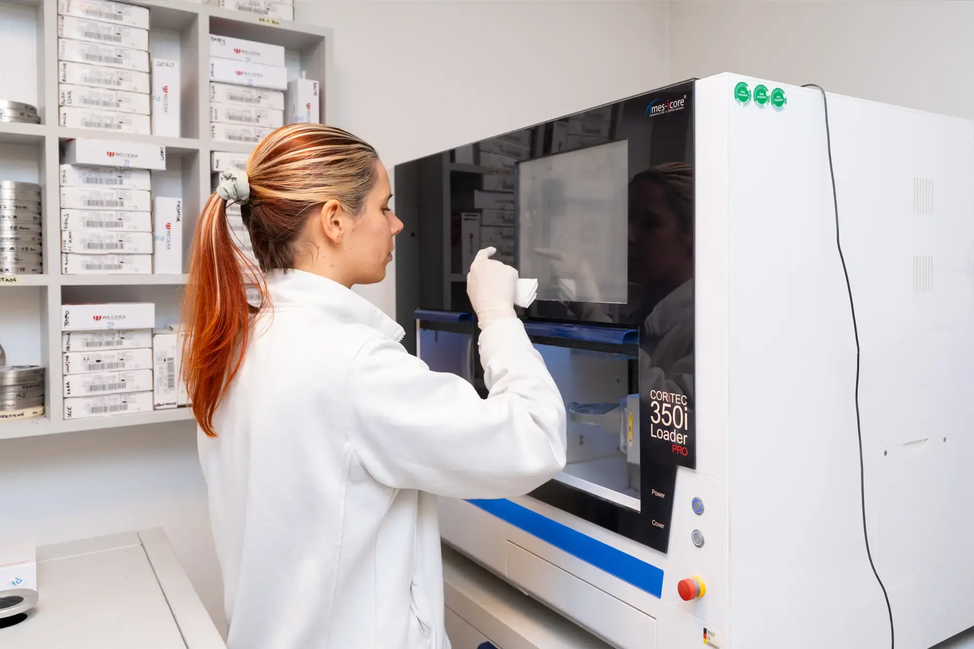
What are they used for?
- Prosthetic restorations – Precision manufacturing of crowns, bridges, veneers, and other restorations with a high degree of accuracy.
- Implantology – Planning and production of custom abutments for dental implants, ensuring optimal fit and functionality.
- Orthodontic therapy – Fabrication of customized orthodontic appliances and aligners tailored to individual patients.
- Surgical guides – Accurate production of guides for implant procedures, enhancing safety and precision.
- Denture production – Digital fabrication of partial and full dentures with optimal fit and aesthetic appeal.
Benefits for users
- High precision and quality - Advanced technology ensures minimal deviations, optimal fit, and long-lasting restorations.
- Increased efficiency and productivity - Automated processes reduce production time and allow for handling more cases in less time.
- Material flexibility - Supports a wide range of materials including zirconia, titanium, PMMA, wax, and composites.
- System integration - Open architecture allows seamless integration with various CAD/CAM software and scanners.
- Cost reduction - Streamlined processes and reduced need for outsourcing result in significant savings.
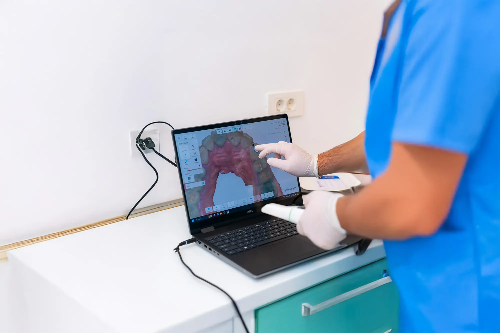
3Shape system in dentistry
What is the 3Shape system?
3Shape is a leading provider of advanced digital solutions for the dental industry. Their portfolio includes intraoral scanners and CAD/CAM software that enable dentists and dental technicians to perform accurate diagnostics, treatment planning, and the creation of dental restorations. This technology significantly enhances service quality and patient satisfaction.
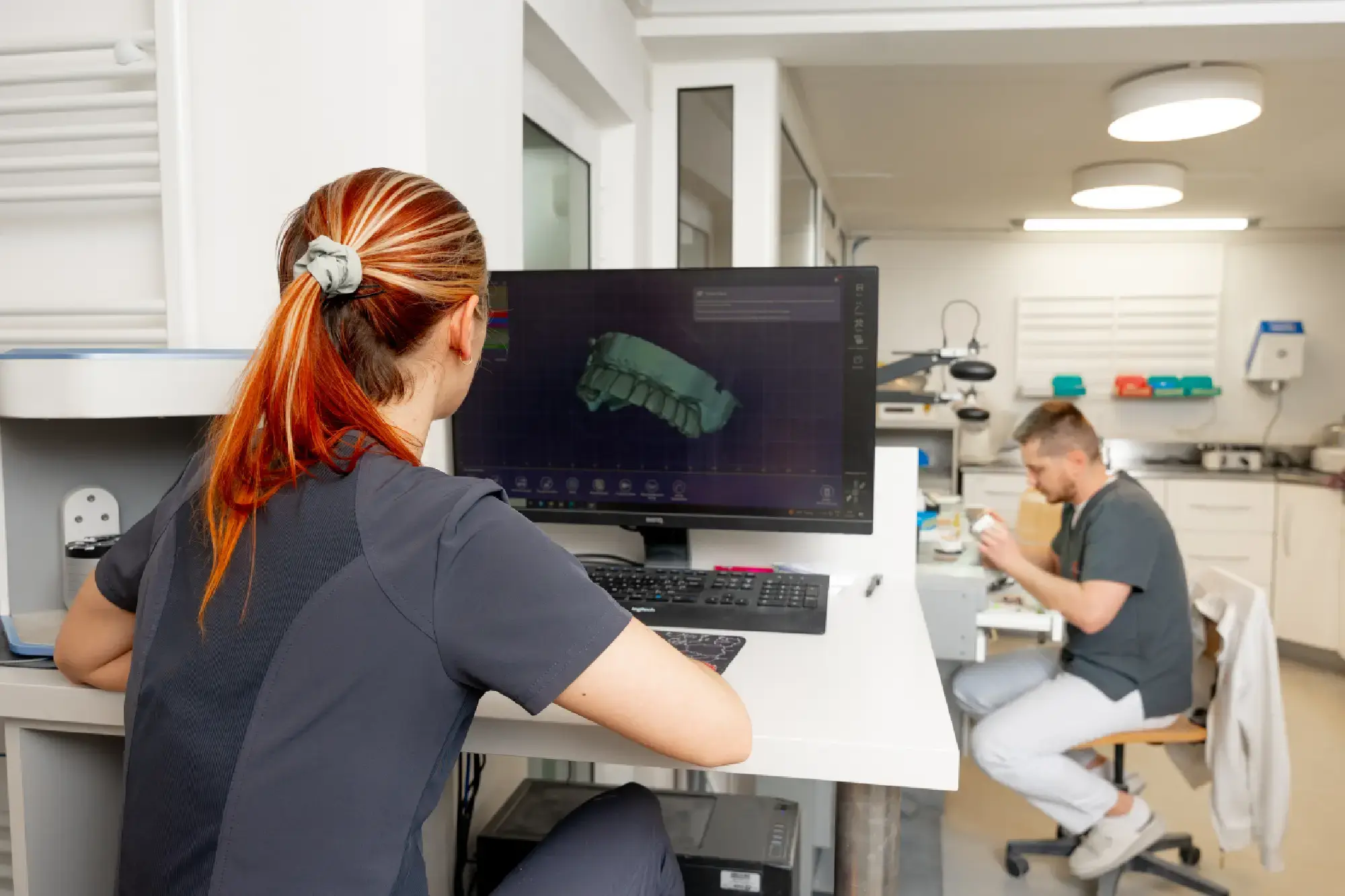
Applications of the 3Shape system
- Intraoral scanning - 3Shape TRIOS scanners allow fast and accurate digital impressions, eliminating the need for traditional methods and increasing patient comfort.
- Designing dental restorations - With the 3Shape Dental System software, labs can design crowns, bridges, veneers, and other restorations with high precision, ensuring perfect fit and aesthetics.
- Orthodontics - 3Shape’s orthodontic software enables detailed planning and fabrication of braces and clear aligners, offering more effective and visually appealing treatments.
- Implantology - Integration with CBCT imaging allows for precise planning and placement of dental implants, leading to safer and more predictable procedures.
- Digital smile design - Advanced software allows for virtual tooth repositioning and smile simulations before starting treatment, helping patients better understand the proposed plan.
Advantages for patients and dentists
- Greater precision and quality - Digital impressions and designs reduce errors, resulting in better-fitting and longer-lasting restorations.
- Faster procedures - Digital workflows reduce turnaround time, minimizing the number of appointments needed.
- Improved communication - Digital data improves collaboration between clinics and dental labs, ensuring efficient and accurate results.
- Increased patient comfort - Eliminating traditional impressions reduces discomfort and improves the overall experience.
- Visualization of results - Patients can preview simulations of their future smiles, boosting confidence and satisfaction with the treatment.
Prosthetic materials in dentistry
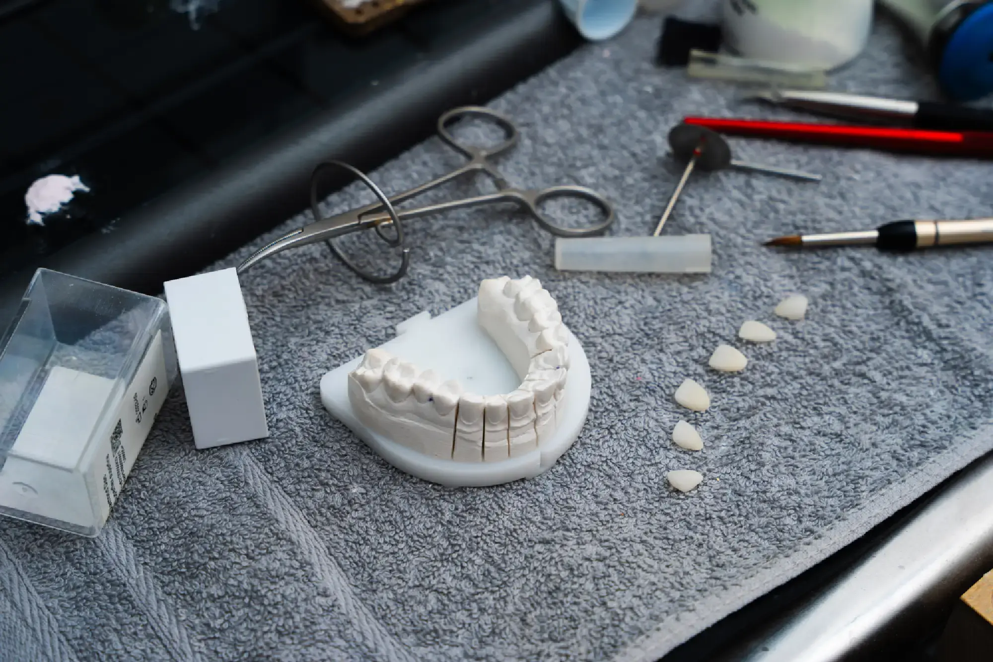
Types of prosthetic materials
- Metal-ceramic– Metal-ceramic crowns and bridges consist of ametal substructure coated with ceramic. This combination offers thestrength of metal and the aesthetic appeal of ceramic. However, overtime, gum recession may expose a dark margin at the junction, butdespite this, metal-ceramics remain a reliable and durable option.
- Zirconia-ceramic – Zirconia is a high-quality ceramic material known for its strength and biocompatibility. Zirconia crowns and bridges offer excellent aesthetics, natural translucency, and long-lasting results. Unlike metal-ceramic, zirconia does not cause dark lines or gum discoloration, making it ideal for the aesthetic zone.
- Lithium disilicate ceramic (E-max) – E-max ceramics are known for their outstanding aesthetics and translucency, enabling the fabrication of restorations that closely resemble natural teeth. This material offers sufficient strength for both anterior and posterior regions while maintaining a natural look.
- Metal alloys – Metal alloys such as gold or titanium are valued for their durability and biocompatibility. Though less aesthetic, these materials are extremely long-lasting and are often used in posterior restorations where appearance is not a primary concern.
- Acrylics and composites – These materials are frequently used for temporary prostheses or removable dentures. Acrylics are lightweight and easy to process, while composites provide improved strength and appearance but are less durable compared to ceramics or metals.
Factors influencing material choice
- Aesthetics - For anterior teeth, where appearance is paramount, materials like zirconia and E-max are preferred for their natural translucency and color.
- Strength and Durability - Posterior teeth endure higher chewing forces, making metal-ceramic or strong metal alloys a more suitable choice.
- Biocompatibility - Materials must be compatible with oral tissues to minimize the risk of allergies or irritation.
- Cost - Financial considerations are also important, as some materials come with a higher price due to superior properties or aesthetic benefits.
Contact us
Our friendly team will be happy to listen to your requests.

